Top stories


Marketing & MediaWarner Bros. was “nice to have” but not at any price, says Netflix
Karabo Ledwaba 21 hours


Logistics & TransportMaersk reroutes sailings around Africa amid Red Sea constraints
Louise Rasmussen 14 hours

More news


New findings about a protein called the nogo receptor are offering fresh ways to think about keeping the brain sharp.
Scientists have found that reducing the nogo receptor in the brain results in stronger brain signalling in mice, effectively boosting signal strength between the synapses, the connections between nerve cells in the brain. The ability to enhance such connections is central to the brain's ability to rewire, a process that happens constantly as we learn and remember. The findings are in the March 12 issue of the Journal of Neuroscience.
The work ties together several research threads that touch upon the health benefits of exercise. While those benefits are broadly recognized, how the gains accrue at a molecular level has been largely unknown. The new research gives scientists a way to produce changes in the brain that mirror those brought about by exercise, by reducing the effect of the nogo receptor.
The find comes as a surprise, because for much of the last decade, the nogo receptor has been a prime target of researchers trying to coax nerves in the spinal cord to grow again. They named the protein after its ability to stop neurons from growing. Its action in the brain has not been a hot topic of study.
The find by neuroscientists at the University of Rochester Medical Centre casts the nogo receptor in a new light. Instead of serving as a target for efforts at regrowing spinal nerve fibres – indeed, the Rochester team showed last year that the molecule doesn't control that process – the molecule suddenly has much broader implications for learning and memory.
“One of the central questions in neuroscience is – what is the molecular and cellular basis of learning?” said Roman Giger, Ph.D., associate professor in the Department of Biomedical Genetics, who led the study. “The nogo receptor seems to play a role.”
The receptor is a promiscuous molecule that hooks up with several other molecules that prevent the growth of neurons in the spinal cord. For most of this decade, scientists have worked to target the molecule, thinking that if they could block it, they could possibly regenerate nerves, repairing spinal cord damage in a way that is impossible today.
However, that road has proved difficult. Last year in the same journal, the Rochester team led by Giger showed that while the nogo receptor does play a role in preventing spinal nerves from growing, it does not control the process outright. While nogo receptor activation can transiently stunt the growth of neurons, it is not required for chronic outgrowth inhibition of injured nerve cells.
Giger's team has found that in some areas of the brain, such as the hippocampus, the nogo receptor is at least 10 times more prevalent than in the spinal cord.
In the brain, Giger's team found that the nogo receptor wields broad influence over a process known as neuroplasticity, which describes how our brain cells change and adapt constantly to meet our needs. It can be thought of simply as the brain's ability to rewire itself on the fly to meet the demands of an organism. The process explains why people are able to recover many of their abilities even after a traumatic brain injury or a stroke: Other brain cells pick up the work for the ones that have died.
Giger's team found that the nogo receptor plays an important role in changing the brain in two ways.
First, the molecule plays a completely unexpected role manipulating the strength of signals between brain cells in the synapses. A team led by Peter Shrager, Ph.D., professor of Neurobiology and Anatomy, made sophisticated measurements of the strengths of the signals as they passed from cell to cell in mice. They found that mutant mice with fewer nogo receptors than normal had stronger brain signalling, what scientists call “long-term potentiation.”
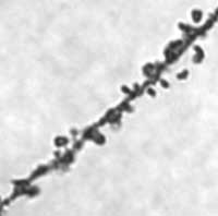
The molecule also affected tiny structures known as dendritic spines, crucial connections that are extensions of the neuron and help cells “talk” to other cells. Mice with lots of the nogo receptor had a different mix of dendritic spines than normal mice. In the hippocampus, the mutant mice had fewer mushroom-shaped dendritic spines and more stubby and thin spines than the other mice. Scientists don't yet know the ramifications of the change, but they say it's firm evidence that the nogo receptor has effects on the anatomic structure of the brain. Creation and removal of dendritic spines is an important form of brain rewiring.
The team attributes much of the effects of the nogo receptor to its ability to strongly bind to a growth factor known as FGF2 (fibroblast growth factor 2), which in the brain and other parts of the central nervous system nourishes neurons, allowing them to branch out and grow new sprouts. When the nogo receptor is present in abundance, it binds to FGF2 molecules, and as a result, neurons no longer branch and sprout as they otherwise would.
Altogether, the findings show that the nogo receptor has a broad impact on processes in the brain that underlie learning and memory, said Giger.
“It's known that changes in synaptic strength can lead to rewiring of the nervous system in such a way that we can compensate for mild to moderate injuries,” said Giger, who is a scientist in the Centre for Neural Development and Disease. “Enhancing synaptic plasticity can partially counter the effects of an injury like stroke, or traumatic brain injury. Really, the process happens routinely in many stroke patients – it's what makes rehabilitation after stroke possible.”
Much of the same type of rewiring also happens as a result of exercise. Scientists have shown that exercise improves the brain's neuroplasticity, boosting the brain's ability to sprout new structures and send crisp signals, which in turn helps people recover from injuries to the central nervous system. Recently, researchers at the Karolinska Institute in Stockholm showed that exercise reduces the abundance of the nogo receptor in the brain. Giger's work provides a molecular framework that brings the disparate findings together.
The findings could also explain something that has puzzled scientists, said Giger. Mice with damaged spinal cords that have been treated with compounds designed to knock out the nogo receptor seem to improve a bit, even though scientists have never been able to demonstrate nerve regrowth in those mice. It may be that their improvement instead is coming through the signal-boosting effect in the synapses.
While it's tempting to think that knocking down the nogo receptor is a simple process that would help people under all circumstances by boosting their brain power, Giger points out that the molecule is not only found at synapses but also along axons, where scientists believe it plays an important role limiting the sprouting of nerve fibres. Any effort to reduce the nogo receptor will have to be studied thoroughly to watch for other effects.
The National Institute of Neurological Disorders and Stroke, the New York State Spinal Cord Injury Research Program, and the Dr. Miriam and Sheldon G. Adelson Research Medical Foundation's Adelson Program in Neural Repair and Rehabilitation funded the work.
While Giger headed the project, much of the research was done in equal part by the two first authors, Research Assistant Professor Hakjoo Lee, Ph.D., and graduate student Stephen Raiker.
Other authors include former graduate student Karthik Venkatesh, Ph.D., now at the University of Michigan; former Professor Hermes Yeh, Ph.D., now at Dartmouth; technician Rebecca Geary; graduate student Laurie Robak; and Yu Zhang, Ph.D., now a research assistant professor in the Department of Neurosurgery.