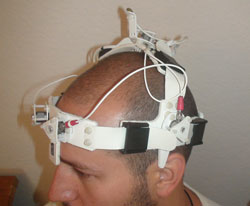
A medical device developed by retired U.S. Navy sonar experts, using submarine technology, is a new paradigm for the detection, diagnosis and monitoring of stroke, says a team of interventional radiologists at the Society of Interventional Radiology's 36th Annual Scientific Meeting in Chicago.
Each type of stroke and brain trauma is detected, identified and located using a simple headset and portable laptop-based console. The device's portability and speed of initial diagnosis (under a couple of minutes) make it appropriate for many uses outside of the hospital setting, including by military doctors in theatre who need to assess situations quickly and efficiently in order to provide critically injured troops with treatment.
"We have developed and validated this portable device for the detection of stroke that is based on decades of submarine warfare technology," said Kieran J. Murphy, M.D., FSIR, professor and vice chair, director of research and deputy chief of radiology at the University of Toronto and University Health Network, Toronto, Ontario, Canada. He said this technology could easily differentiate normal brain from life-threatening conditions, such as swelling (haematoma) and bleeding (haemorrhage). "For example, when a physician suspects stroke time is of the essence, doctors could use the system to determine treatment that needs to begin immediately," he added.
The device's continuous monitoring capability - unique in neurodiagnostics - will allow immediate detection of changes in a patient's condition. In addition to being ideal for field ER, ambulance or military use, the researchers' hope is for the technology to be adapted and used in other areas of acute care, such as open heart surgery (where stroke is an ever-present concern), in other vascular indications elsewhere in the body and in monitoring the progression of disease for drug efficacy.
A deadly condition
"Stroke is the third leading cause of death in the United States and the leading cause of disability," explained Murphy. A stroke or "brain attack" occurs when a blood clot blocks an artery or a blood vessel breaks, interrupting blood flow to an area of the brain, he said. When either of these things happens, brain cells begin to die and brain damage occurs. And, stresses Murphy, "time to diagnosis is critical to improving patient outcomes; time is brain."
"The system is very simple in principle, yet it yields exceedingly rich data," said Murphy. He explained that the device's basis in submarine technology means it works to measure a patient's complex brain pulsations and to provide information on the type and location of an abnormality in many of the same ways as sonar works on submarines. Both use an array of sensors to measure movement and generate signals to be processed and analysed, matching the signals to objects or conditions. "As sonar sorts out whales and other objects from vessels, the device sorts out cerebral abnormalities such as aneurysms, arteriovenous malformations (AVMs, an abnormal connection between veins and arteries), ischemic strokes and traumatic brain injury from normal variations in physiology," noted Murphy.

Traditionally in transcranial ultrasound, the thinner areas of the skull are used as "windows" through which ultrasound can "see" or "listen," said Murphy. This system does not rely on these "windows." It measures the movement of the skull (acceleration) to separate confounding acoustic signals from the motion created by the in-rushing blood flow in this way: blood flows into the brain during each pulse and a pressure wave emulates from the vessels outward in all directions. The wave encounters the skull and accelerates it. The device measures the acceleration and records this complex waveform with a time synchronised high-resolution digitiser, explained Murphy.
Gold-standard imaging
In a 40-patient proof-of-principle trial, the patients - 16 men and 24 women - with a wide variety of cerebrovascular conditions, including intracerebral haemorrhage, subarachnoid haemorrhage, intracranial aneurysms, AVMs, ischemic stroke and transient ischemic attack (an episode in which a person has stroke-like symptoms for up to 1-2 hours), were studied. The researchers employed gold standard imaging - using CT magnetic resonance imaging or catheter angiography - to verify the diagnosis of the patients, who were then measured. The analysis team was blinded as to a patient's clinical history. For normal controls the research team relied on data taken on some 30 normal subjects (apart from the study). The team's algorithms were able to separate normal from all other conditions, to separate all patients with a specific condition into their own category and to provide information about the location of the abnormality.
Initial blood vessel models were used to identify data signatures that were subsequently verified on patients in the clinic. The interventional radiologists said that, as they continue to increase their signature library, their expectation is that the location capability, as well as the capacity to detect other neurologic conditions with a high degree of sensitivity and specificity, will also increase.
More information about the Society of Interventional Radiology, interventional radiologists and minimally invasive treatments for many conditions can be found online at www.SIRweb.org.
Abstract 114: "Submarine-based Technology for the Detection of Acute Intracranial Abnormalities and Stroke: Validation in 40 Patients," E.A. Fiona Aldrich, department of medical imaging, University of Toronto, P. Neild, P. Lavoi, research, Jan Medical, Mirimar, California, and K.J. Murphy, radiology, University of Toronto, Ontario, Canada, SIR 36th Annual Scientific Meeting, March 26-31, 2011. This abstract can be found at www.SIRmeeting.org.
Source: Society of Interventional Radiology


