World's first non-surgical closure of leaking heart valve
Marianna Cronjé, the 59-year old female patient from Stellenbosch, was born with her heart transposed in a mirror image of what is considered anatomically normal, it is located on the right side of her chest. Because of rheumatic fever at a young age, Cronjé had undergone four open-heart operations over a period of 40 years. Her last valve replacement in 2000 was successful, but due to scar tissue and other difficulties, this was considered her last operation. When this valve developed a leak, she had no surgical options and her condition deteriorated gradually to the point where she could do nothing for herself.
She was referred to Dr Hellmuth Weich, a cardiologist who practices at Mediclinic Panorama and is attached to Stellenbosch University and Tygerberg Hospital, as a candidate for a percutaneous (non-surgical) alternative.
The entire procedure was performed through a needle puncture in the patient's groin. A tube was fed through a vein up into the right heart chamber and then a hole had to be made in the wall of the heart to get across to the left chamber. From here, the leak in the mitral valve had to be crossed and was then closed with two disc-like devices, which are intended for closing birth defects in children's hearts.
"This procedure is technically extremely difficult in a patient with normal anatomy, and has never been done before on someone with dextrocardia," says Weich, who performed the procedure in the catheterisation laboratory of the hospital.
"The six-hour procedure went well thanks to an excellent team of medical professionals. Cronjé is making a slow recovery in hospital." He cautioned that although the procedure was successful, her condition was poor prior to the surgery and that recovery may take a month or more.
Dr Weich will be presenting this case next month to 500 delegates at the World Paediatric and Adult Cardiology Congress in Cape Town.
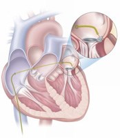
The illustration below indicates how the catheter (thin tube) is inserted in the groin and fed through a vein into the right atrium. From here a needle puncture is made to the left atrium and this tube is fed up to the leaking valve. The zoomed in area indicates how the two devices were positioned next to the leaking valve. It is important to note that this illustration reflects a heart in a normal orientation but that in Cronjé's case it was a mirror-image (dextrocardia with situs inversus).