Medtech News South Africa

Philips launches live 3D image guidance tool for tumours

Philips has launched its latest innovation in interventional oncology, the EmboGuide, a live 3D image guidance tool to treat difficult-to-reach tumours or tumours in patients who are deemed unsuitable for surgery.
Philips launches live 3D image guidance tool for tumours

It is designed for use in conjunction with the company's interventional X-ray system to perform tumour embolization procedures. Such procedures involve blocking the arteries feeding a tumour to deprive it of nutrients and oxygen. They require the insertion of a catheter, which must be guided to the tumour site with the aid of live image-guidance.

Developed in collaboration with leading clinicians and partners such as BTG (btgplc.com), an innovator in interventional oncology, it addresses the need for an enhanced 3D imaging solution to make interventional oncology procedures more effective and easier to perform and ultimately improve patient outcomes. It offers interventional radiologists the ability to visualise, characterise tumour lesions and plan and execute interventional procedures.

"Interventional oncology is a fast growing field that offers clinicians a viable treatment option for patients who are not suitable for surgical tumour removal," says Jose Fernandes, Philips Healthcare MD for Southern Africa. "Together with our partners, we will leverage our combined expertise in image-guided interventions and therapies to accelerate this transformation from surgical procedures to minimally-invasive treatments in oncology."

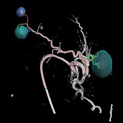
A specific example of a tumour embolization procedure is trans-arterial chemo-embolization (TACE), used for palliative treatment of liver tumours. It involves simultaneous local administration of chemotherapy and beads that block the arteries feeding the liver tumour.

We can only treat what we see, yet the embolization of all blood vessels that feed the liver tumour lesion is key for an effective TACE procedure. The live 3D image guidance helps to improve the technical success of the procedure, as it can automatically identify the small tumour-feeders that are difficult to detect with conventional 2D angiographic imaging methods.

Advantages

  • See - it leverages the ultra-low X-ray dose settings of the AlluraClarity interventional X-ray system and the fast, high quality imaging of the abdomen of XperCT Dual. XperCT Dual's enhanced imaging technique (multiphase cone beam CT) offers high quality 3D images of lesions, with proven detection accuracy superior to conventional 2D angiographic imaging. It offers clinicians a better view of the treatment targets for informed decision making while performing the procedure. XperCT Dual is comparable to MRI, which is considered to be the 'gold standard'.
  • Reach - it helps to define the tumour lesions and features automatic identification of blood vessels that feed the lesions. It detects more than twice as many tumour-feeding arteries compared to conventional imaging methods (Digital Subtraction Angiography (DSA). This allows interventional radiologists to optimise the catheter locations for embolization and plan a route to them. During the administration of the embolization agent, it accurately superimposes the planning information onto the interventional X-ray system's live images to monitor the treatment progress and determine its endpoint.
  • Treat - "In the treatment of intermediate HCC, the integrated use of our embolic device with image guidance is vital for successful treatment," said Mike Motion, BTG GM, Interventional Oncology. "As a leader in interventional oncology, we invest to improve the safety and efficacy of the procedure. Partnering with Philips has strengthened our ability to further develop this cohesive approach using DC Bead and EmboGuide with the ultimate objective of advancing interventional oncology."

Let's do Biz